Updated Report-Based Impression

Formal MRI interpretation now drives the conclusion

The principal diagnostic source in this repository is now the formal MRI report electronically signed on March 13, 2026. That report supports prior anterior instability with extensive anteroinferior labral tear, osseous Bankart, a large intra-articular body in the axillary recess, and an on-track Hill-Sachs deformity.

It also explicitly states that there is no rotator cuff tear and no subacromial-subdeltoid bursal fluid or contrast leak. The report also describes torn labral tissue and likely glenoid osseous fragment migrating to the axillary recess with surrounding proliferative change, resulting in the large intra-articular body.

The X-ray arthrogram PDF remains useful for procedure confirmation only. It documents successful intra-articular contrast injection before MRI and does not provide the main diagnostic conclusion.

Main finding
Anterior instability pattern with extensive labral tear and osseous Bankart
Also present
Large loose body in the axillary recess
Associated injury
On-track Hill-Sachs deformity with edema
Explicitly not seen
Rotator cuff tear or bursal contrast leak

Report Summary

Updated interpretation after review of the MRI report

The formal MRI report is now the principal interpretive source in this repo. The findings below summarize that report and cross-reference it to the available study images.

Main problemHigher confidence

Anterior instability with labral tearing and osseous Bankart

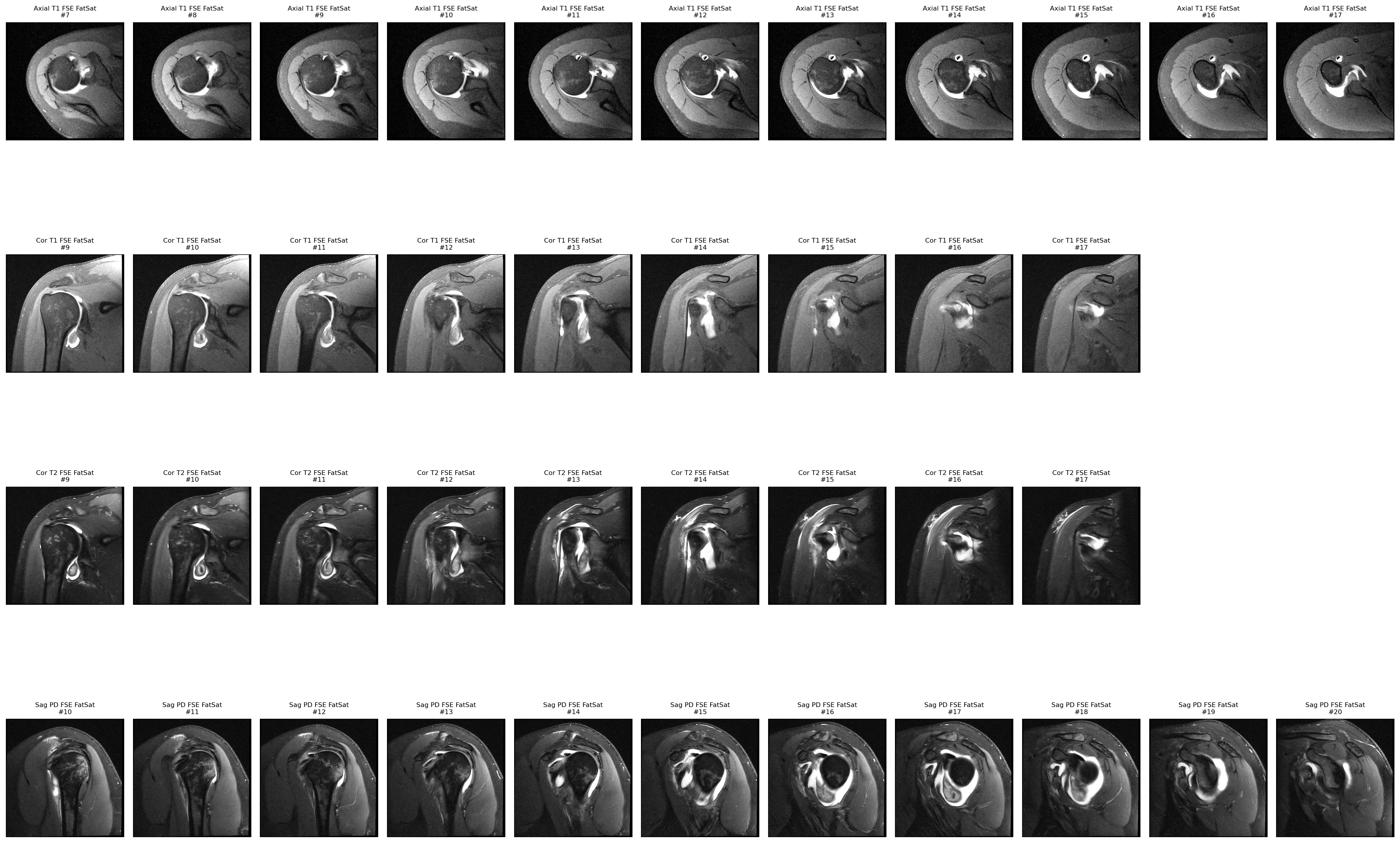
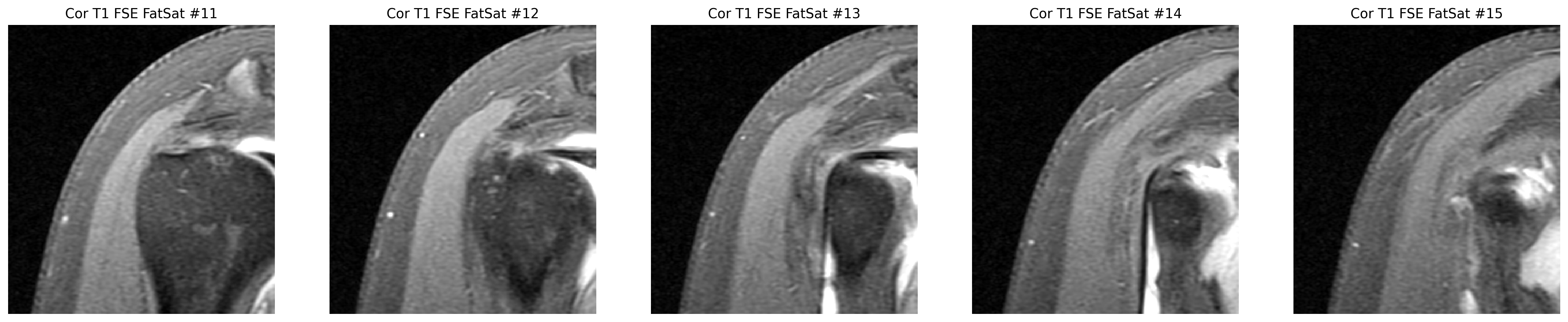
The formal MRI report identifies an anterior instability pattern with extensive anteroinferior labral tearing and osseous Bankart.

Coronal MRI slices from the right shoulder arthrogram.
The formal MRI report centers the case on anterior instability, labral tearing, and osseous Bankart. These coronal arthrogram slices are part of that broader study context.

Supporting points

  • The MRI report says the anterior inferior labrum is torn from at least 3 to 6 o'clock.
  • It also says the labrum is absent in the anterior superior quadrant and is more likely torn than simply a normal variant.
  • It further describes abrupt truncation of the anteroinferior glenoid from 4 to 6 o'clock, in keeping with osseous Bankart.

Cine viewer

Right Shoulder Image Review

StudyMRI series loading

Study type

Pick MRI or X-ray

Series selection

MRI series loading
Loading stack viewer…

Full preview board

Loading study

Browse all images

Every derived study image, in a cleaner image browser

The browser is now built around the selected image first, with a large preview, category-aware navigation, and grouped thumbnails so it is easier to understand what you are looking at.

Overview1 of 1

Key series overview

Composite panel of the most useful sequences.

Open full size
Visible now1images in the current filter
Category total1images in Overview
Filekey_series_focus.pngoriginal derived image filename
Key series overview

Selected image

Overview image from the derived study set.

1 / 1

Overview

Search and filter on the right, then step through the current result set from here.

Navigator

Search by filename or series, then browse by category.

1 of 25 images

Overview

Composite summary panels across the study.

1

Terms translator

Search the medical words

Arthrogram

An MRI done after contrast is injected into the joint to make small internal tears easier to see.

Labrum

A ring of cartilage around the shoulder socket that helps stabilize the joint.

Instability

A shoulder that has partly slipped or dislocated and is now prone to doing it again.

Loose body

A fragment of tissue or bone floating inside the joint space.

Hill-Sachs deformity

A dent or compression injury on the humeral head that commonly happens after anterior shoulder dislocation.

Glenoid

The socket side of the shoulder joint that the humeral head sits against.

Rotator cuff

The group of shoulder tendons that help lift and rotate the arm. In this report, those tendons are described as not torn.

Method and limits

How this site was built

Used as evidence

Raw DICOM image series, exported PNG image panels, the fluoroscopy PDF, and the formal MRI report PDF signed on March 13, 2026.

Explicitly not used

The old README, outside internet sources, and any clinic notes or records that are not actually present in this repo.

Main limitation

The site is now anchored to the formal MRI report, but it is still a patient explainer rather than a treatment recommendation.

Step 1

Load the study and the report

7 MRI series, 1 fluoroscopy series, and the MRI report signed on March 13, 2026 were reviewed directly from the repo assets.

Step 2

Cross-check the report on the images

The report's main calls were checked against the sagittal, coronal, and axial series in the repo.

Step 3

Final synthesis

Once the formal MRI report was present, the site was revised so the primary summary reflects instability, extensive labral tear, osseous Bankart, large intra-articular body, and Hill-Sachs change.